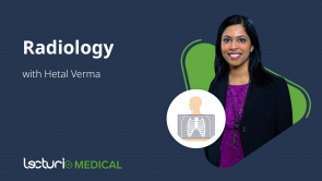
Chest CT Technique and Anatomy

Über den Vortrag
Der Vortrag „Chest CT Technique and Anatomy“ von Hetal Verma, MD ist Bestandteil des Kurses „Thoracic Radiology“.
Quiz zum Vortrag
What would be appropriate Hounsfield units of a lipoma on CT?
- -100 to -50
- 0
- + 50 to +100
- -1000
- +500
Which of the following statements is FALSE?
- Radiation is multiplicative, so multiple scans should be done whenever possible.
- The radiation dose varies based on the type of machine, type of scan, and the patient’s body size.
- The Hounsfield unit for water is 0.
- Density is the amount of radiation that a structure absorbs.
- Window levels are the digital manipulation of the image to accentuate structures of various Hounsfield units.
Which plane of a CT image looks at the patient from the feet up to the head?
- Axial
- Sagittal
- Coronal
- Oblique
- Parasagittal
All of the following structures are seen in a coronal CT section of the mediastinum EXCEPT?
- Left minor fissure
- Right atrium
- Aorta
- Left ventricle
- Pulmonary artery
Kundenrezensionen
5,0 von 5 Sternen
| 5 Sterne |
|
5 |
| 4 Sterne |
|
0 |
| 3 Sterne |
|
0 |
| 2 Sterne |
|
0 |
| 1 Stern |
|
0 |


